




| IN A NUTSHELL |
|
Repeated exposure to shock waves, such as those experienced by military personnel, can have profound and lasting effects on brain function. Recent research led by Harvard Medical School has uncovered evidence that these impacts may not be visible on standard brain scans, yet still significantly affect brain connectivity and functionality. This study, which focused on U.S. special operations forces, has revealed how trauma from bomb blasts increases the risk of traumatic brain injury (TBI) over time. The findings highlight the need for more refined diagnostic tools to identify and address these hidden injuries, which often manifest as severe symptoms in affected individuals.
Understanding Blast-Induced Brain Trauma
Military personnel frequently face the risk of exposure to blast waves during their service, which can lead to significant brain trauma. The study conducted by Harvard Medical School researchers focused on the special operations forces within the U.S. military. They aimed to understand how repeated exposure to such blasts could lead to traumatic brain injuries, even when traditional brain imaging techniques fail to show any visible damage.
Key findings highlighted that service members with high levels of blast exposure exhibited significant differences in functional connectivity within their brains. This refers to how various regions of the brain communicate and function together. The study linked these alterations with more severe neuropsychological symptoms, including memory issues and emotional difficulties. As neuroradiologist Andrea Diociasi noted, “In short, repeated trauma seems to weaken the brain’s internal communication.”
Invisible Injuries and New Diagnostic Approaches
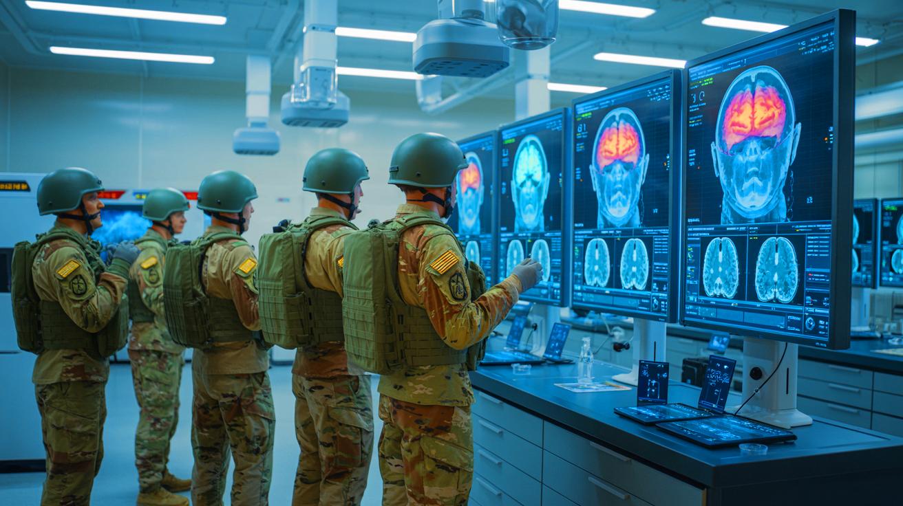
One of the primary challenges in diagnosing and treating brain injuries in military personnel is that many injuries are “invisible” on standard scans. The research team conducted advanced MRI analyses with greater detail, using statistical models to detect changes that are typically overlooked. Their efforts resulted in a predictive model capable of identifying brains exposed to high blast levels with 73 percent accuracy.
Diociasi also pointed out that certain brain regions appeared larger in individuals with greater blast exposure, indicating possible long-term tissue changes such as scarring. “These aren’t injuries you can always see with the naked eye, but they are real – and now we can start measuring them,” she stated. This novel approach opens up possibilities for earlier detection and intervention, changing how brain injuries are assessed.
Broader Implications for Brain Injuries
The researchers believe that their findings are not limited to military-related brain injuries. The study’s methodologies and insights could be applied to other forms of brain trauma, such as those resulting from contact sports or serious workplace accidents. By mapping how trauma leads to changes in brain connectivity, the research provides a framework for understanding the clinical symptoms that follow.
This comprehensive map of trauma’s effects on the brain could lead to better assessments and treatments. “The findings reveal that even when the brain looks normal, it might still be carrying hidden signs of trauma – and we now have tools to detect them,” Diociasi explained. Such advancements offer hope for more effective interventions and a deeper understanding of repeated trauma’s long-term effects on the brain.
Future Directions and Potential Treatments
The implications of this research extend beyond diagnosis to potential treatment strategies. By identifying specific changes in brain connectivity associated with trauma, new therapeutic targets may be developed to mitigate the effects of repeated exposure to shock waves. The predictive model and detailed brain maps could guide personalized treatment plans, offering hope for improved quality of life for affected individuals.
The study’s success in identifying hidden brain injuries underscores the need for ongoing research and innovation in the field of neuroimaging. As technology continues to evolve, so too does the potential for earlier detection and more effective management of traumatic brain injuries. The publication of these findings in the journal Radiology marks a significant step forward in understanding and addressing the silent injuries that impact countless individuals.
As we continue to delve into the complexities of brain injuries resulting from repeated trauma, the question remains: How can these new insights be translated into practical solutions for those affected by such invisible wounds?